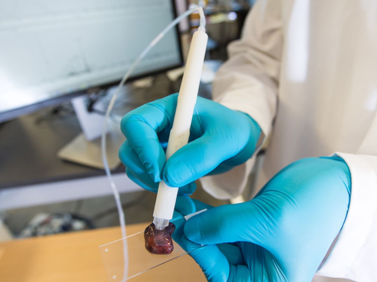
Scientists have invented a gentle, handheld device that can determine within seconds whether tissue is cancerous or not. The tool could give surgeons precise information during an operation about which tissues should be removed or preserved.
The pen-shaped instrument, which relies on the chemical analysis technique mass spectrometry, was developed by scientists at the University of Texas at Austin. It is described in a report published today in the journal Science Translational Medicine.
“The presence of a tumor after surgery is strongly associated with cancer recurrence and decreased survival,” says Livia Eberlin, associate professor of chemistry at the University of Texas at Austin who led the development of the pen. “So it is best to remove it all the first time.”
Getting every last bit of cancerous tissue is difficult. The trouble spots usually occur at the boundary of the tumor, where it’s hard to tell whether the tissue is benign or cancerous. Breast cancer patients often undergo second and third surgeries to remove cancerous tissue left behind after the initial procedure.
But removing too much benign tissue around the margin of the tumor can affect how well an organ functions. That leaves surgeons constantly making judgment calls during operations about what to snip.
The current tool for testing tissue during an operation, called frozen section analysis, involves cutting, freezing, and staining a slice of tissue, and then having a pathologist visually inspect it. The process takes at least 30 minutes, and can lengthen an operation. Freezing tissue can also change its structure, resulting in inaccurate results.
Eberlin’s “MasSpec” pen could provide a diagnosis right away without a pathology lab. Her team tested it on 253 human tissue samples and found that it correctly identified cancerous tissue more than 96 percent of the time.
The handheld instrument itself is simple, but it interfaces with something far more complex: a mass spectrometer. These are large machines that identify molecules based on their masses and the trajectories they take through an electric or magnetic field. Eberlin’s pen functions as an attachment to a mass spectrometer.
The system works like this: A surgeon holds the pen against the questionable tissue. Suspended at the tip of the pen is a tiny drop of water, and when it comes into contact with the tissue, any water-soluble molecules on the tissue, including cancer-specific metabolites, naturally dissolve into the water droplet.
The droplet is then pumped through tubing to the mass spectrometer, which provides information on each of the molecules present in the sample—information such as the molecules’ mass-to-charge ratios, which is the quantitative relation between a molecule’s mass and electrical charge, and is unique for each molecule.
That information is then analyzed by a statistical classifier called Lasso, an algorithm developed 20 years ago. Eberlin’s team trained the algorithm on hundreds of samplesto recognize cancer biomarkers based on their mass-to-charge ratios.
Within ten seconds, the tool displays the result: either “normal” or “cancer”. A surgeon would likely need to take samples from multiple spots around the margin of the tumor. Eberlin is working on software that would spatially map the results.
This is not the first time engineers have proposed a tool that can identify cancer in real-time during surgery. Scientists have experimented with other mass spectrometry–based tools, along with fluorescent probes, and different forms of spectroscopy. But all of them, in the process of analyzing the tissue, damage it in some way, because they require the use of gas, solvents, voltages, or heat.
Eberlin’s MasSpec pen, which uses only water, causes no damage to the tissue. And the instrument is 3D-printed with a cheap, disposable tip.
But there’s a practical problem: The system requires a mass spectrometer in the operating room. “They’re not common at all” in hospitals, Eberlin says. “That’s definitely the roadblock to translating this.”
Mass spectrometers are expensive—easily half a million dollars—and are usually only found in high-tech research, forensic, and drug detection labs. Eberlin, for her experiments, used a commercially available mass spectrometer from ThermoFisher Scientific that is the size of a ‘90s-era copy machine.
Eberlin says she envisions developing a smaller, cheaper mass-spec machine with only the necessary capabilities for her diagnostic pen. “It could be an instrument that is rolled in and out of surgery rooms,” she says.
Indeed, other researchers earlier this year described improvements to mass spectrometry technology that make it cheaper, more portable, and more sensitive than ever before.
Eberlin plans to initiate next year a pilot study in which surgeons will test her pen’s diagnostic capabilities during actual surgeries. She’ll compare the results with the current clinical approaches to cancer patient care, she says.
Emily Waltz is a features editor at Spectrum covering power and energy. Prior to joining the staff in January 2024, Emily spent 18 years as a freelance journalist covering biotechnology, primarily for the Nature research journals and Spectrum. Her work has also appeared in Scientific American, Discover, Outside, and the New York Times. Emily has a master's degree from Columbia University Graduate School of Journalism and an undergraduate degree from Vanderbilt University. With every word she writes, Emily strives to say something true and useful. She posts on Twitter/X @EmWaltz and her portfolio can be found on her website.


