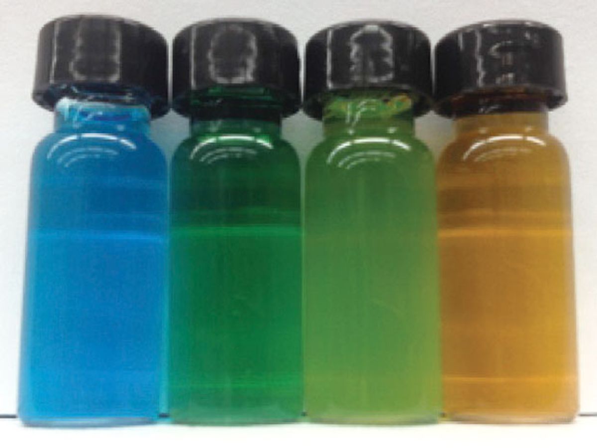
Researchers at the University of Buffalo (UB) have developed what they're calling a "nanojuice", which when ingested enables doctors to see clear images of the small intestine in real time.
The novel medical imaging technique promises better diagnosis of a variety of gastrointestinal illnesses, including Crohn’s disease and celiac disease. Other medical imaging techniques used to examine the small intestine, such as X-rays, magnetic resonance imaging, and ultrasound, have drawbacks in terms of safety, accessibility to the organ, and an inability to produce clear images.
Perhaps the biggest breakthrough of this technique is that unlike other imaging techniques it is capable of monitoring what’s happening in the small intestine in real time.
“Conventional imaging methods show the organ and blockages, but this method allows you to see how the small intestine operates in real time,” said Jonathan Lovell, assistant professor of biomedical engineering at UB in a press release. “Better imaging will improve our understanding of these diseases and allow doctors to more effectively care for people suffering from them.”
The key to the technique is the ingestion of a liquid with nanoparticles suspended in it, thus the name "nanojuice." The basis of the nanoparticles is a family of dyes known as napthalcyanines. While these molecules are great for absorbing light that make them ideal as a contrasting agent, alone they are unsuitable for use in the human body. First, they don’t disperse in a liquid; and, secondly, they could be absorbed in the intestine and transferred into the blood stream.
To counteract this, the UB researchers developed nanoparticles they dubbed “nanonaps” that contain the dye molecules inside them, imparting the ability to both disperse in liquid and pass through the intestine without problems.
In the research, which was published in the journal Nature Nanotechnology, the UB team gave the nanojuice to mice orally and then used photoacoustic tomography—a kind of ultrasound imaging that uses light-induced pressure waves. The result was that nanoparticles in the intestine could be visualized with low background and a high resolution.
This technique enables for the first time the visualization of peristalsis, which involves the contraction of muscles that moves food through the small intestine. The ability to observe this process in patients could not only help in the diagnosis of gastrointestinal illnesses but also help determine the link between peristalsis dysfunction and ranges of disorders, including diabetes and Parkinson’s disease.
The researchers plan to take this work to the next step, human trials, and test the technique in other areas of the gastrointestinal tract.
Dexter Johnson is a contributing editor at IEEE Spectrum, with a focus on nanotechnology.


