The compound eyes of an insect are structurally very different from that of a human—almost a separate category of organ altogether from the more familiar ocular arrangement of lens, cornea, iris, and retina. For instance, Papillio xuthus, the Asian Swallowtail Butterfly, can see wavelengths of light in the ultraviolet spectrum, far beyond the limits of human vision or the capabilities of standard optical electronics in cameras and mobile phones.
Researchers at the University of Illinois Urbana-Champaign (UIUC), inspired by P. xuthus’ unique eyes, have developed a new kind of camera sensor that can detect ultraviolet light. Recently published in Science Advances, the new device extends the capabilities of a real-time optical sensor with a layer of fluorescent crystal that makes it sensitive to ultraviolet light (UV). This design, which mimics the function of fluorescent compounds in the eyes of P. xuthus, may enable the development of new medical devices and imaging technologies.
“As an engineer, you always wonder: what principles can we mimic from [nature’s] visual systems to make better cameras?”
—Viktor Gruev, University of Illinois Urbana-Champaign
The xuthus butterfly can see UV light because of two complementary mechanisms. One is a set of cells in their eyes that, much like the shortest cone cells in human eyes, are most sensitive to blue light but are also partially sensitive to near-blue UV light. However, to see shorter wavelengths of light in the UVA and UVB ranges—280 to 400 nm—the butterfly uses a clever trick: cells containing fluorescent dyes within the insect’s eyes radiate a band of green light when excited by UV radiation. These dyes translate otherwise invisible UV light into a signal that the butterfly can see.
Viktor Gruev, a professor at UIUC and an author of the present study, says that the idea to mimic P. xuthus’ visual system in silico resulted from years of collaboration between him and biologists studying how different animals see. “I’ve been working with visual biologists for over ten years,” says Gruev, whose previous collaborations have produced other bio-inspired cameras. “As an engineer, you always wonder: what principles can we mimic from those visual systems to make better cameras?”
The UIUC team built the sensor using a layered CMOS design with a top layer of fluorescent perovskite crystal. Similar to a Foveon chip, the three photosensitive layers of the sensor divide the spectrum of visible light into three bands with peak sensitivities at blue, green, and red. These bands are determined by the width and position of each layer, with red light penetrating down to the lowest sensor layer and blue light only activating the highest layer.
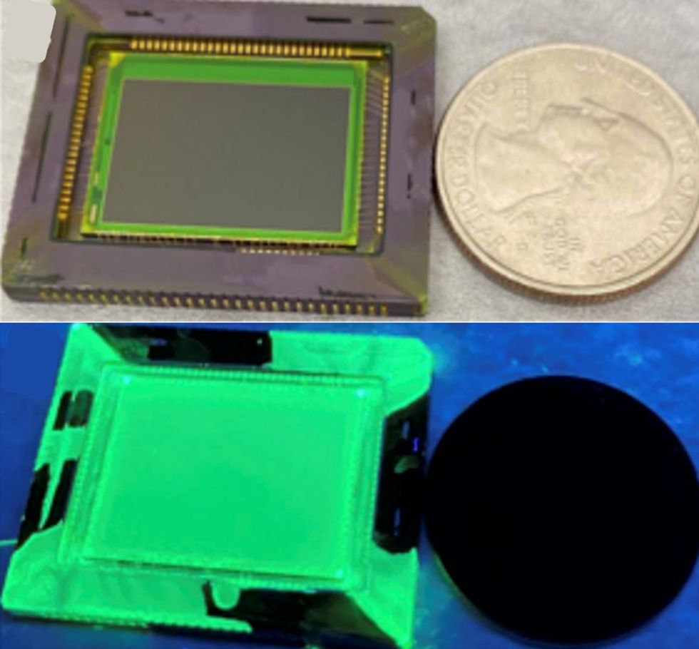 The UV imaging sensor is seen here under white light [top] and under UV light [bottom]. The green appearance is attributed to PNC layer fluorescence.University of Illinois Urbana-Champaign/Science Advances
While this CMOS sensor arrangement does mimic the partial UV sensitivity of the layered photoreceptor cells in xuthus’ eyes, the real magic in the UIUC chip comes from a layer of perovskite crystal deposited on the sensor’s surface. The perovskite nanocrystal (CsPbBr3) is both fluorescent under UV radiation and transparent to visible light. This fluorescence causes the crystal layer to glow green when irradiated with UV light.
Just like the fluorescent dyes found in P. xuthus eyes, the perovskite nanocrystals translate incoming UV light into a wavelength that the CMOS sensor already responds to. Paired with the partial sensitivity of the blue-band sensor layer to near-UV light, the perovskite fluorescence enables the sensor to resolve exact wavelengths of UV light—as small as 250 nm—in real time and at any point on its surface. “The UV light is detected in two components,” says Shuming Nie, a UIUC professor and another author on the paper. “The first is the UV absorbed by the nanocrystals that convert it into a visible signal—in our case, green—and that’s picked up by the photodiode. There’s also a residue UV picked up by the top layer photodiode. We can use the ratio of those two to calculate the UV wavelength.”
The researchers say this is a big step forward in UV sensing technology. “There is no, that I’m aware of, UV sensing technology that gives you a 2D image in real time with exact wavelength resolution all at once,” says Gruev. “UV spectrometers can give you more spectral information, but only at a single point.”
The researchers propose that their fast, flexible sensor has direct biomedical applications. Cancer cells overproduce certain kinds of proteins and amino acids fluoresce in the UV spectrum when irradiated. Clinical devices can use this principle to detect cancerous tissues quickly and directly. The researchers show in the paper that the device can resolve the distinct fluorescent UV signatures of the cancer-indicating proteins and amino acids tyrosine, elastin, tryptophan, and nicotinamide adenine dinucleotide (NADH). They also show that the device can distinguish between lab cultures of cancerous and non-cancerous cells based on their UV fluorescence.
In future work, the researchers plan to expand on their design by adding multiple perovskite crystal layers that fluoresce in different bands, which would provide better UV wavelength resolution. “A better way to resolve wavelengths is with two-color nanocrystals,” says Nie. “This gives you more real estate, more space, to resolve the UV wavelengths.” Adding more color-tuned crystal layers would heighten the device’s cancer detection capabilities, Nie says. “With only one layer of nanocrystal,” he continues, “Some of the UV signals from amino acids in tumor cells are clustered together. If we use the two-color crystals, they are better separated.”
- One-Eyed Bug Vision Helps Drones Land - IEEE Spectrum ›
- Mantis Shrimp Eyes Inspire Cameras to See Cancer - IEEE Spectrum ›
- Self-Driven “Vine” Seeks Light and Heat - IEEE Spectrum ›