“Brain Age” AI Discovers Early Alzheimer’s Signs
Improved detection rates plus increased saliency could equal hope
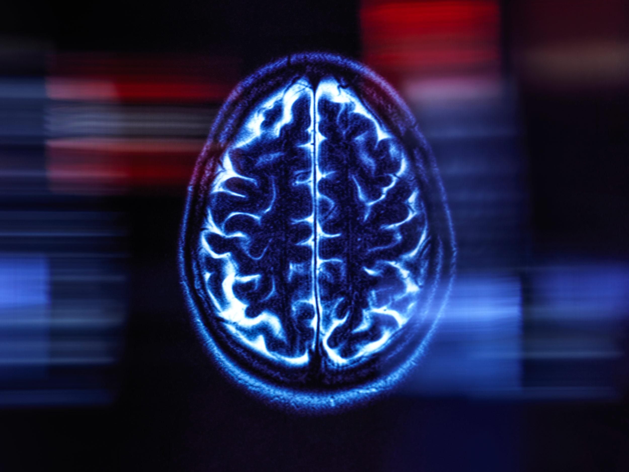
Although your brain has chronologically the same number of years in the world as the rest of you, determining its medical “age,” a measure of how processes of aging impact the brain, is far from obvious or straightforward. Now, researchers are testing an AI-based model that determines brain age via magnetic resonance imaging (MRI) data, potentially helping identify early signs of dementia and Alzheimer’s disease.
This latest research is hardly the first machine-learning model to be applied to the “brain age” problem. But the new research, published in the Proceedings of the National Academy of Sciences, looks like it could help buy high-risk patients crucial time.
“If we do identify people at high risk early, we can mitigate the risk through lifestyle changes or potential treatments,” said Andrei Irimia, an assistant professor of gerontology at the University of Southern California and the senior author of the study.
Irimia and his colleagues trained their model using MRIs from 4,681 cognitively normal patients sampled from a combination of databases, with the largest number coming from the UK Biobank. The researchers used a different set of 1,170 MRIs from the same databases to test their network. For each MRI, the neural network produces an estimate of a patient’s chronological age. The researchers referred to this value as the patient’s brain age, which should be as close to a person’s chronological age as possible when it is given scans from cognitively normal adults.
The algorithm predicted chronological age with an average error of about 2.3 years, which the researchers said is more accurate by about a year than other comparable brain-age techniques.
“I think their improvement is meaningful,” said Han Peng, who was previously a postdoctoral researcher at Oxford University and helped develop a similar neural network to determine brain age that previously was the most accurate.
Of course, as with much of AI, brain-age algorithms are often black boxes—inscrutable from inspection and unrevealing about how they generated their findings. Irimia’s team, however, wanted to make their algorithm interpretable. So it also generates what are called saliency maps—showing the areas of the MRIs the network relies most on to make its decisions.
The researchers used 650 more MRIs from cognitively normal patients, as well as 359 who had Alzheimer’s dementia and 351 with less severe effects from the disease called mild cognitive impairment (MCI). About half of the patients with MCI later developed dementia. The saliency maps supported findings from other studies, including ones on normal aging in the brain and how Alzheimer’s might impact the brains of male and female people differently.
“We found that our method could confirm and reproduce other findings from other studies that use completely different methods,” Irimia said.
The researchers found that a larger gap between brain age and chronological age increased the risk that someone with MCI would eventually develop dementia, which previous studies have also found. They also found that for patients with either MCI or dementia, brain age was more correlated with their level of cognitive functioning than their chronological age was. This was also true of patients with MCI, but not for the group with dementia. That may be because the model was trained using data from cognitively normal patients, the researchers write, although there could be multiple explanations, Irimia said.
Though this study used a large data set, the sample still skews heavily toward white people of European descent, said James Cole, a professor of neuroimage computing at University College London. Specific racial demographics are not given for the study because the authors didn’t have access to that information, said Irimia. However, the UK Biobank, the study’s largest data source, is about 95 percent white. It’s crucial that researchers be able to show that their research applies to a diverse group of people, said Cole.
It’s hard to say how meaningful the one-year increase in accuracy is, said Eran Dayan, an associate professor in the department of radiology at the University of North Carolina, Chapel Hill’s school of medicine.
To know this, Dayan said, future research would have to incorporate more clinical data. Though this study did incorporate some data on cognitive function, Dayan says more studies using longitudinal data, or patient data over time, would be needed to eventually use this technology with real patients.
Irimia doesn’t disagree. “I think we need more research to really understand how these [saliency map] patterns are different and how we could leverage the information we have gained to improve risk assessment,” he said.
- Can Big Data Help Prevent Alzheimer’s Disease? ›
- AI Could Analyze Speech to Help Diagnose Alzheimer’s ›
Rebecca Sohn is a freelance science journalist. Her work has appeared in Live Science, Slate, and Popular Science, among others. She has been an intern at STAT and at CalMatters, as well as a science fellow at Mashable.