Bioprinting is the use of 3D printing techniques to fabricate tissues out of biomaterials. It is mainly used to create human tissue for research and for drug testing in vitro. When used to create a body part intended for implantation in a patient, the part must first be printed with a desktop bioprinter, and then large open-field surgery is typically required to place it. Besides the risk of infection and long recovery time, a mismatch between the printed part and the internal target tissue it’s being attached to is possible, as are problems arising from contamination and handling.
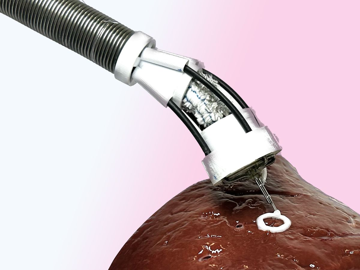
To overcome these challenges, researchers at the University of New South Wales Sydney, in Australia, have developed a miniature soft robotic arm and flexible printing head, and integrated them into a long tubular catheter that makes up the flexible printer body. Both the arm and printing head have three degrees of freedom (DoFs).
“Our flexible 3D bioprinter, designated F3DB, can directly deliver biomaterials onto the target tissue or organs with a minimally invasive approach,” says Thanh Nho Do, a senior lecturer at UNSW’s Graduate School of Biomedical Engineering, who together with his Ph.D. student, Mai Thanh Thai, led the research team.
Not only does F3DB have the potential to directly reconstruct damaged parts of the body, it “can also be used as an all-in-one endoscopic surgical tool with the nozzle taking on the role of a surgical knife,” Do adds. “This would avoid the need for using different tools for cleaning, marking, and incising now used in longer procedures, such as removing a tumor.”
Prototype flexible 3D bioprinter can also serve as an all- purpose endoscopic surgical tool. Source: UNSW Sydneyyoutu.be
Though in situ bioprinting has been investigated for the past decade, “bioprinting onto internal organs has been limited due to various difficulties,” says Ibrahim Ozbolat, professor of engineering science and mechanics at Pennyslvania State University, in commenting on the research details published in February’s Advanced Science. “This mobile all-in-one endoscopic bioprinting device is novel,” he says, and could “advance existing techniques by allowing real-time observations, incisions, and bioprinting onto internal organs.”
The device has a similar diameter to that of an endoscope (about 11 to 13 millimeters), small enough to be inserted into the body through the mouth or anus. The soft robotic arm is actuated by three soft-fabric-bellow actuators regulated by a hydraulic system composed of DC-motor-driven syringes that pump water to the actuators. A flexible printing head, consisting of soft hydraulic artificial muscles, enables the printing nozzle to move in three directions, like that of a conventional desktop 3D printer. Overall control is by a master-slave setup that uses a commercial haptic system to transmit hand motions by the master.
On reaching the target, the arm and printing head are controlled by an automated algorithm based on inverse kinematics, a mathematical process that determines the motions necessary to deliver the biomaterials onto the surface of an internal organ or tissue. Printing is monitored by an attached flexible miniature camera.
To test the device, the researchers first used various nonbiomaterials such as liquid silicone and chocolate to print different multilayer 3D patterns in the lab. In further experiments, they printed various shapes with nonliving materials on the surface of a pig’s kidney. Later, the researchers printed in situ living biomaterials on a glass surface inside an artificial colon.
“We saw the cells grow every day and increase by four times on day seven, the last day of the experiment,” says Do.
To test the device as an all-purpose tool for endoscopic surgery, the researchers performed various functions such as washing, marking, and dissecting the intestine of a pig. “The results show the F3DB has strong potential to be developed into an all-in-one endoscopic tool for endoscopic submucosal dissection procedures,” says Do.
Further improvements are needed, including the inclusion of more parameters in the kinematic model that controls the printing, and the addition of more cameras to better monitor the printing. “Then we will begin testing the device on animals, and eventually on humans,” says Do. “We hope to see the device in operation in hospitals in the next five to seven years.”
The device “has high potential to be successful,” Ozbolat agrees. “But its safety needs to be verified first and other improvements carried out.” He notes that 3D endoscopic robot arms are already in use clinically, so provided that the device’s feasibility and safety is proven going forward, “commercialization can only be a matter of time.”
- 3D Printing - IEEE Spectrum ›
- 3D Printing Bone Directly Into the Body - IEEE Spectrum ›
- A 3D Bioprinter Makes a Spinal Cord Implant in 1.6 Seconds - IEEE ... ›
- Landmark Transplant Turns 3D Bioprinting on Its Ear - IEEE Spectrum ›
- Holograms Supercharge Nanoscale 3D Printing - IEEE Spectrum ›
- Origami Inspires Sensor Implanting Method for Bio-Printing - IEEE Spectrum ›


