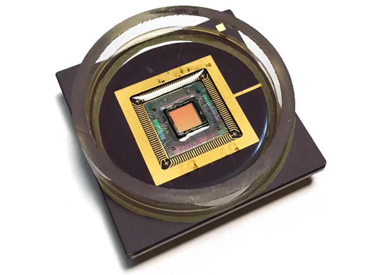
Researchers at Harvard University have developed a nanoelectrode array capable of imaging the electrical signals within the living cells. While other technologies have been able to measure these signals, this new complimentary metal oxide semiconductor nanoelectrode array (called a CNEA) can measure these signal across an entire network of cells.
“It’s similar to an imager combining the light signal from each detector in a pixel array to form a picture; the CNEA combines the electrical signals from within each cell to map the network level electrical activities of the entire cell culture,” explained Donhee Ham, a professor at Harvard involved in the research, in an e-mail interview with IEEE Spectrum.
This network-level intracellular recording capability can be used, for example, to examine the effect of pharmaceuticals on a network of heart muscle tissue, enabling tissue-based screening of drug candidates. It could also help better understand how cells communicate with each other across a network.

In research described in the journal Nature Nanotechnology, the CNEA device fabricated by the Harvard team looks like a normal CMOS integrated circuit, except that there are nanoscale electrodes fabricated on the surface of the chip and a cell culture chamber on top.
The CNEA device contains an array of 1,024 (32×32) pixels with the distance between the center of each pixel to the center of the next one (the pitch) being 126 micrometers. Beside the nanoelectrode, each pixel includes an amplifier to record the electrophysiological events, a stimulator to manipulate the cell membrane voltage, and a memory to change the state of the pixel operation between recording and stimulation.
In operation, the cells are cultured right on top of the chip and are in direct contact with the nanoelectrodes. “The electrical signals from the cells are collected by the nanoelectrodes, amplified and multiplexed on the chip, transferred through a customized printed circuit board in which the chip is plugged into, and are finally read by a data-acquisition card and PC,” said Ham.
The look and operation of the CNEA device are very similar to microelectrode arrays (MEAs) and planar patch-clamp chips, which have been leading the way for how all-electrical electrophysiological imaging has been recorded. However, the CNEA device departs from this two technologies by managing to combine their greatest strengths in one device while eliminating their weaknesses.
MEA’s for example can only record signals from outside the cell. Those signals “do not have the high sensitivity characteristic of intracellular recording,” said Ham. Microfluidic patch-clamp arrays, can record high-precision intracellular signals, but they can’t do so at the level of a network of cells.

There were two big challenges in fabricating the CNEA devices, according to Ham. The primary one was constructing the nanoelectrodes on top of the CMOS chip, which was fabricated in a foundry. The second issue was developing a front-end circuit—the amplifiers etc.—that could interface with such small—thus high-impedance—electrodes.
While Ham and his colleagues were able to overcome these challenges, some key engineering work still remains if the device is to be commercialized. In particular, the fabrication process must be standardized and the electrode/cell interface must be better understood and optimized.
The next version of the system is already in the works: “The upgraded CMOS circuit will have more electrodes and a finer pitch, with better circuit performance and more functionality at the pixel level,” explained Ham. “The optimized nanoelectrodes will improve the signals acquired from cells and will also allow the platform to be used for different types of cells in both the in vitro (cell culture) and ex vivo (tissue slices) systems.”
Dexter Johnson is a contributing editor at IEEE Spectrum, with a focus on nanotechnology.


