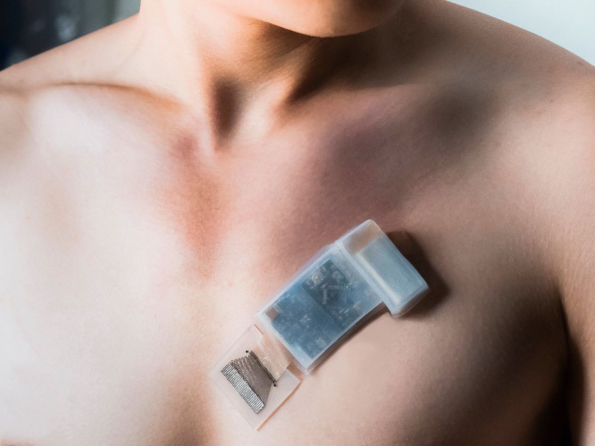
Researchers have made the first wireless wearable ultrasound device that images tissue deep below the skin while patients move around freely. The system could allow doctors to monitor blood pressure, heart health, and lung capacity in real time while the wearer goes about their daily activities, including during workouts or other physical exertion.
“Clinicians want to know how those parameters change, say, with exercise, and they can’t do that now,” says Muyang Lin, a doctoral candidate in neuroengineering at the University of California, San Diego. “This kind of system has always been a dream for a lot of companies making ultrasound probes. But this is the first demonstration of such a system.”
The credit-card-size ultrasound patch can monitor signals from tissues as deep as 16 centimeters under the skin. It can continuously measure blood pressure, heart output, respiratory health, and other physiological signals for up to 12 hours on a single charge.
“We’re not aiming to achieve very deep imaging. We’re more interested in motion sensing, because that’s the advantage of the wearable system for continuous monitoring.”
—Muyang Lin, University of California, San Diego
Traditional ultrasound imaging works by bouncing high-frequency sound waves off tissue and analyzing the echoes received by the ultrasound probe to reconstruct images. And while it is safer and less expensive than other imaging techniques like MRI, conventional ultrasound relies on bulky equipment and skillful technicians to operate the probes. “Manipulation of the ultrasound probe plays a key role in collecting crisp images and reliable information,” Lin says. “So ultrasound now always requires manual operation.”
This also means that the images cannot be collected continuously. To monitor cardiac health, for instance, ultrasound imaging is done before and after vigorous exercise. But that misses vital information on the heart as it pumps blood during exercise.
A few research groups are working on wearable ultrasound systems. But all previous devices reported to date have been tethered to other hardware and wired up to an external power source and data-collection device, says Sheng Xu, a neuroengineering professor at UCSD and coauthor of a paper the team published in Nature Biotechnology. Xu’s group reported on—and IEEESpectrum previously reported on—one such wearable cardiac imager just a few months ago.
The new system is the first that is fully integrated. It consists of a stretchable ultrasonic probe made of a 32-channel array of piezoelectric transducers—converting electrical signals to sound waves and vice versa—that transmit and receive ultrasound signals to and from the body.
This probe is connected to a flexible control circuit via serpentine copper wires that are stretchable. The control circuit drives the transducers to generate ultrasound pulses and receive the echoes. The echo signals are amplified and sent to a microcontroller unit that converts them to digital signals, which a Wi-Fi transmitter sends to a computer or smartphone. A commercial lithium-polymer battery powers the circuit.
The brains of the system consist of a machine-learning algorithm that the team developed to analyze the signals from the ultrasound patch. By measuring the pulsation of the carotid artery and the jugular vein, the system calculates the wearer’s heart rate and blood pressure. It can also measure the contraction of the heart muscle to estimate the volume of blood the heart pumps out, and monitor the motion of the diaphragm to calculate respiratory output.
The device was able to continuously monitor these signals even when a participant biked outside for 30 minutes and performed vigorous high-intensity interval training exercises. When the patch moves relative to the tissue underneath, the algorithm selects the best channel in real time, Lin explains, producing data continuously during the wearer’s motion. “As long as one of the 32 channels is receiving, the algorithm decides in real time which one gives the best signal.”
Conventional ultrasound has its place, and this wearable won’t be replacing it any time soon, the researchers say. The signal quality is limited as is the distance that the system can peer into the body. But the ability to continuously monitor vital signals 12 hours at a time on moving patients has “a lot of clinical value,” Lin says. “We’re not aiming to achieve very deep imaging. We’re more interested in motion sensing, because that’s the advantage of the wearable system for continuous monitoring. We want to enable on-body monitoring with a wireless system, so people can go anywhere and do anything while those signals are monitored.”
- Ultrasound Stickers Look Inside the Body ›
- Wearable Ultrasound Patch Images the Heart in Real Time ›
- How MEMS Ultrasound Imaging became Ultra Small - IEEE Spectrum ›
Prachi Patel is a freelance journalist based in Pittsburgh. She writes about energy, biotechnology, materials science, nanotechnology, and computing.


