Many of us are acquainted with magnetic resonance imaging (MRI), but you may not have heard of nanoMRI a technology that can image viruses and cells for in-depth analysis.
Up until now, nanoMRI has been a long and laborious process, in which a single scan can take up to two weeks to complete. Now researchers at the Swiss Federal Institute of Technology in Zurich (ETH Zurich) have dramatically reduced this scanning time to a couple of days by developing a system that can perform in parallel the measurements that used to have to be done sequentially—a process known as multiplexing.
"As a loose analogy, think of how your eyes register green, red, and blue information at the same time using different receptors—you're measuring different colors in parallel," Alexander Eichler, a postdoctoral researcher in the physics department at ETH Zurich said in a press release.
Fundamentally, MRI exploits the fact that particular atoms have nuclei that are like tiny magnets. When these atoms come under the influence of a magnetic field, they start to rotate around the axis of that magnetic field.
This rotation is called precession. The precession generates its own frequency of electromagnetic radiation known as the Larmor frequency. The Larmor frequency depends on the type of atoms and the strength of the magnetic field. So an MRI determines the positions of the atoms based on frequencies of their precession.
This standard MRI process works pretty well until you get down to nanoscale objects like cells where the strength of around 1000 atoms is not robust enough to be detected.
"Clinical MRI is only possible because a single 3-D pixel—a ‘voxel’—contains about 1018 atoms," Eichler explained in the press release. "With nanoMRI, we want to detect voxels with only a thousand atoms or less, meaning that we need a sensitivity at least a quadrillion [1015, or a million billion] times better."
In the research published in the journal Applied Physics Letters, the ETH Zurich team developed their technique around a magnetic resonance force microscopy (MRFM) apparatus, which has been the standard piece of equipment for previous nanoMRI methods.
With the MRFM apparatus, the nuclei of the atoms are exposed to a small magnetic force that is transferred to a micro-scale cantilever. The force on the cantilever causes it to vibrate, and that vibration can then be measured in a way that forms an image.
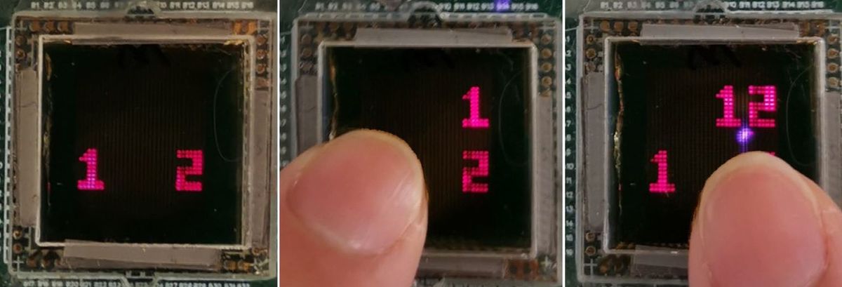
This process used to have to be done one measurement at a time but the ETH researchers have developed a technique to do make these measurements in parallel across six data points. This parallel measurement, known as multiplexing, involves encoding different bits of information in the detector using different phases.
"The term 'phase' refers to a lag in a periodic signal,” said Eichler. “The phase can be used to differentiate between periodic signals in a way similar to how color is used to differentiate between light signals in the eye."
Eichler added: “With commercial applications in mind, this time gain is crucial because it makes a huge difference to a pharmaceutical company if a virus can be characterized within three days rather than a month."
Dexter Johnson is a contributing editor at IEEE Spectrum, with a focus on nanotechnology.