Researchers at Cardiff University in Wales have developed a new method for taking readings of processes going on inside living cells. The technique, which relies on nanodiamonds, could eventually aid in developing new modes of drug delivery and cancer therapeutics.
The traditional method for imaging cellular processes has depended on fluorophores, which are a fluorescent chemical compound that can re-emit light upon light excitation. But they degrade under the light used to illuminate them. This renders them ineffective as visualization targets after a short period of time. Furthermore, fluorophores have been shown to become toxic over time, sometimes killing nearby cells.
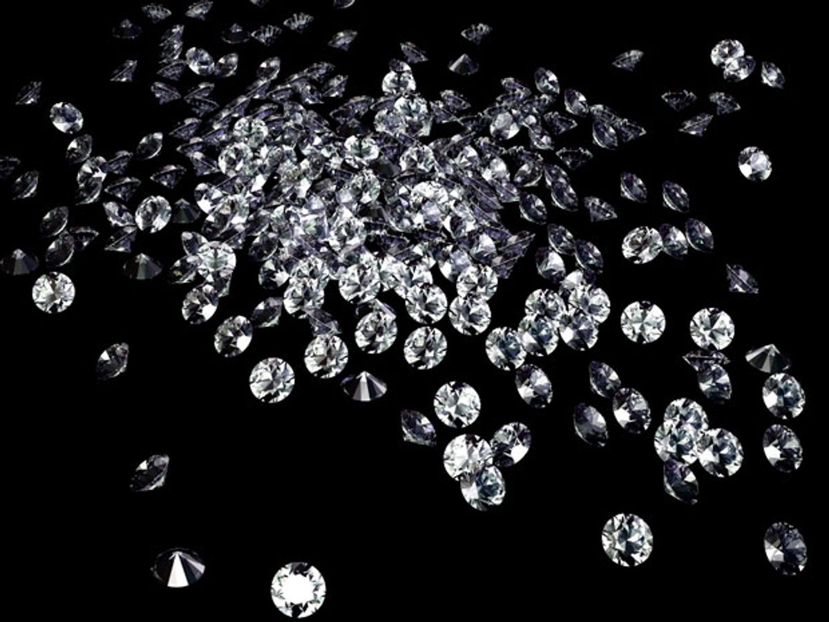
Nanodiamonds have been proposed as a one-to-one replacement for fluorophores for sometime now. But getting them to fluoresce required designing small defects into them. Manufacturing these defects into the diamonds proved to be costly and time consuming.
In research published in the journal Nature Nanotechnology, the Cardiff researchers developed a new method wherein the nanodiamonds are used differently, thus eliminating the need for this difficult trick of producing them with specific defects.
Instead, the Cardiff team demonstrated that nanodiamonds without defects can be imaged optically through the interaction between the illuminating light and the vibration of the chemical bonds inside the diamond lattice structure. These vibrations cause the light to scatter in such a way that they produce a different color.
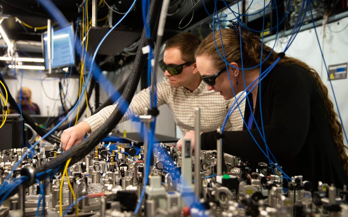
To get this effect, the researchers used two laser beams pulsing at a specific frequency so that laser light triggers the chemical bonds inside the nanodiamonds to vibrate in sync. The researchers then focus one of two lasers at these vibrations, which produces a light known as, coherent anti-Stokes Raman scattering (CARS).
The researchers were then able to use a microscope to measure the intensity of the CARS light on a series of nanodiamonds of varying sizes. After using electron microscopy and other optical contrast methods developed by the researchers, the team was able to accurately measure the sizes of the diamonds. This then made it possible to quantify the relationship between the size of the nanodiamonds and the intensity of light that they produce.
The end result was a method that allowed the researchers to measure the size and number of nanodiamonds that had been delivered into the living cells.
“This new imaging modality opens the exciting prospect of following complex cellular trafficking pathways quantitatively with important applications in drug delivery,” said Paola Borri from the School of Biosciences, who led the study, in a press release. “The next step for us will be to push the technique to detect nanodiamonds of even smaller sizes than what we have shown so far and to demonstrate a specific application in drug delivery.”
Dexter Johnson is a contributing editor at IEEE Spectrum, with a focus on nanotechnology.


