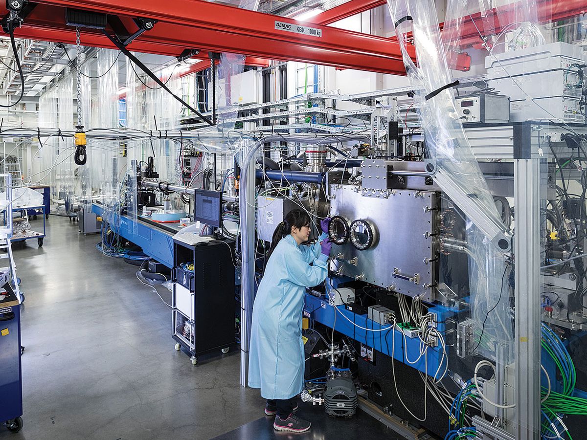
The European XFEL, or EuXFEL—currently the largest X-ray free-electron laser in the world—produced its first light in May 2017. The facility, built in a 3.4-kilometer tunnel near Hamburg, Germany, was commissioned in September of last year. And August 2018 brought another milestone—the publication of the first scientific paper based on experiments with the laser, which the authors used to probe the 3D structures of protein microcrystals.
More results are sure to come as the laser ramps up to full operation. Already, more than 500 researchers from 20 countries have visited the facility to investigate structures of crystalline materials, biomolecules, viruses, and chemical reactions. Two experimental stations are now available to the scientific community, but by 2019, the facility should have six stations supplied with pulses from three X-ray lasers. The cooling requirements of the current design limit it to delivering no more than 27,000 pulses per second, but researchers envisage increasing this to a continuous stream of pulses.
In the EuXFEL, electrons first move through a linear accelerator, mounted in a 2.1-km tunnel and packed with superconducting cavities. Radio waves create alternating positive and negative electric fields within these cavities. The electrons are bunched together so that they enter each cavity at the moment the electric field is positive. An electron injected in the first cavity will accelerate through to the second cavity. Each new cavity gives the electrons a boost of energy.
LCLS-II
The LCLS (Linac Coherent Light Source), at Stanford University’s SLAC National Accelerator Laboratory, was the world’s first laser to produce X-ray pulses, which lasted only femtoseconds. LCLS came on line in 2012, and with 120 pulses per second, it was the most powerful instrument of its kind until the advent of the European XFEL in 2017. But in 2020, the European X-ray laser will itself be surpassed by the LCLS-II, LCLS’s successor. Its capabilities are expected to open up a new era in the research of chemical reactions, biochemistry, materials science, and solid-state physics.
A closer look at LCLS-II:
- Generates 1 million pulses per second
- Produces an X-ray laser beam 10,000 times as bright as that of the LCLS
- Extends the tunable photon energy range to 25 kiloelectron volts
- First operation is expected in the second half of 2020
The electrons emerge from the accelerator with an energy of up to 17.5 gigaelectron volts (GeV) and are injected into three so-called undulators made up of a series of permanent magnets. The magnets have alternating polarities that force the electrons to follow a curvy, sinusoidal path as they pass through the gaps between the magnets’ poles. Each time an electron is deflected from its path, it emits radiation. The X-ray waves produced at each magnet then propagate forward and combine with waves from all the other magnets to produce a coherent, needle-sharp beam of pulses.
The superconducting cavities allow the accelerator to accelerate up to 27,000 bunches of electrons at a time, which produce the same number of X-ray pulses in the undulators—and generate an extremely high-resolution beam. Because so many pulses are produced in such quick succession, researchers can follow chemical reactions and molecular interactions in real time.
“The high repetition rate is our stronghold,” says EuXFEL managing director Robert Feidenhans’l. The EuXFEL generates more data for each experiment than any other X-ray free-electron laser, such as the U.S. Department of Energy’s Linac Coherent Light Source (LCLS), at Stanford University. This makes EuXFEL better suited to mapping small, complex biological structures such as proteins.
For example, a molecule can form a diffraction pattern when blasted by an X-ray pulse, but the pulse destroys the molecule. So to study the molecule’s structure, scientists must continuously present new, undamaged molecules to the pulses by embedding them in a stream of liquid that crosses the path of the X-ray.
This approach was used by researchers from Europe, Australia, and the United States, who published that first paper about proteins in August. They mixed urease, an enzyme present in plant seeds and animal tissues, and two other proteins, found in jack beans, into liquid and sprayed it across the path of the laser. The pulses kept hitting new microcrystals, and collecting diffraction patterns from many of them allowed the researchers to reconstruct the jack bean proteins’ crystalline structures.
This approach could also allow scientists to create “molecular movies” that show how atoms behave during chemical reactions by recording the motion of individual atoms, or to study the behavior of viruses present in a jet of liquid.
The high repetition rate of the EuXFEL also speeds up experiments and permits many more researchers to use the facility, says Claudiu Stan, a physicist at Rutgers University and coauthor of the microcrystals paper. Indeed, the LCLS delivers 120 pulses per second, and the interval between pulses is 8 milliseconds. The EuXFEL can produce 1 million pulses per second, although it cannot deliver more than 27,000 pulses in rapid bursts. During those bursts, the separation between pulses is 222 nanoseconds.
Anton Barty, a researcher at the Center for Free-Electron Laser Science, in Hamburg, led the very first experiment at EuXFEL in September 2017, which supplied the data for a second paper based on research with the laser. “There was so much new technology involved, from the accelerator to the detector, that we were convinced something would go wrong. Yet amazingly, we got publishable results from that very first week,” he recalls.
In addition to exploring protein structures, the EuXFEL will help investigators study the composition of matter under extreme conditions, and how it behaves when exposed to extremely strong magnetic fields. “It would be very interesting to investigate superconducting and electronic materials under these conditions,” Feidenhans’l says.
This article appears in the November 2018 print issue as “Europe’s New X-Ray Laser Delivers Results.”
