A new implant that can be draped over the brain like plastic wrap has successfully been used to simultaneously monitor the activity of broad swaths of the brain and individual neurons, proving effective in early trials with human patients on the operating table. Researchers report in Science Advances that the new tech, called the NeuroGrid, could improve treatment and surgery for epilepsy patients.
The CDC’s 2013 estimates state that about 2.9 million people in the United States have active epilepsy—a seizure-inducing brain disorder. About one third of those people have drug-resistant epilepsy, according to the Johns Hopkins Medicine Health Library. In these cases, doctors can only try to cut out parts of the brain where seizures begin, but it’s challenging to pinpoint the targets.
To prepare for surgery, doctors typically keep their patients in the hospital to monitor suspected parts of their brains for several days or weeks. Often, this monitoring process requires opening up the skull and implanting an electrode grid that fits over the surface of the brain. However, this electrocorticography (ECoG) technology has previously only recorded broad patterns of activity from regions of the brain. Getting precise recordings from individual brain cells was thought to only be possible with penetrating electrodes that are inserted into the brain tissue.
György Buzsáki, a neuroscientist at NYU Langone Medical Center and a coauthor of the new paper, have been working on a better technology for years. In 2015 research published in Nature Neuroscience, he and his colleagues created an electrode grid that’s as clingy as plastic wrap, and that can monitor the activity of both single neurons and groups of neurons. In that prior study, researchers tested the NeuroGrid in rodents. The new study tried out the tech in humans for the first time: Neurosurgeons temporarily placed the implants in five people undergoing brain surgery to measure the technology’s performance.
Buzsáki says this first-in-humans trial is a big step forward. While the brains of rodents and humans are fundamentally the same in some ways—“electricity is electricity,” Buzsáki says—proving a new technology to be safe and reliable requires careful testing. “It doesn’t matter how sure you are in anything—you still have to test it in a patient,” he says.
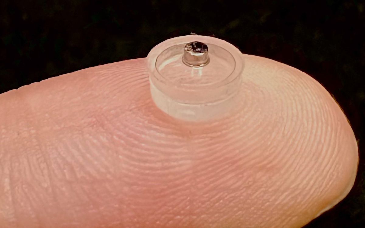
The NeuroGrid is a 4-micrometer-thick sheet made of a polymer that’s already approved for use in the human body. It has 120 conductive polymer electrodes, and can cover about 420 mm2 of the neocortex. It has very similar properties to cellophane wrapping paper, Buzsáki says, because it sticks to wet surfaces such as the brain.
For every 10 electrodes in the NeuroGrid there is a single wire that connects to the lone silicon chip outside the brain that acts as an amplifier. The chip compresses the signals and sends them via wire to a computer for analysis. With this setup, researchers found that they could successfully detect when individual neurons fired, which gave them more precise information about activity in the brain.
Buzsáki says that detecting the activity of an individual neuron is like picking out an individual instrument that’s playing in an orchestra. “Hopefully, recognizing any pathological activity will be easier,” he says.
Study coauthor Dion Khodagholy, who’s a researcher in Buzsáki’s lab, says the highly localized performance comes from a combination of how nicely parylene interfaces with the brain, how nicely the conductive polymer tracks electricity, and the silicon-based microfabrication process.
Mikhail Lebedev, a neuroscientist at Duke University who wasn’t involved in the study, agrees that the tech could “allow [us] to localize the epileptic focus more accurately.” He also sees potential applications relating to his own research, which involves recording brain activity and using it to control prosthetics.
“It’s not instant panacea for a clinical practice,” cautions Nitish Thakor, a neuroengineer at Johns Hopkins University who also wasn’t involved in the study. He often works on brain-draping ECoG arrays that track neuron activity for such applications as the control of robotic limbs.
There are the usual multi-year regulatory requirements for getting such a technology approved for clinical use, Thakor says. Then, although the benefits from recording individual neuron firing are clear, Thakor says the tradeoff is that the new technology would require more electrodes and more sophisticated data processing. “It will pave the way for clinical studies,” he says, “but also offers new challenges.”
Buzsáki says his team’s next goals are to decrease the NeuroGrid’s size and the number of wires coming out, so doctors can use more grids and get even better data about the surgical areas. They may also work on making the power supply and any amplifiers fully implantable inside the skull.