Want to understand what happens to the brain as it ages, or figure out how people learn to recognize faces? Neurologists asking such questions, or struggling to deal with brain degeneration caused by Parkinson’s and Alzheimer’s, might get some insight from detailed observations of the brain’s circuitry over time. But so far, such information has been hard to come by.
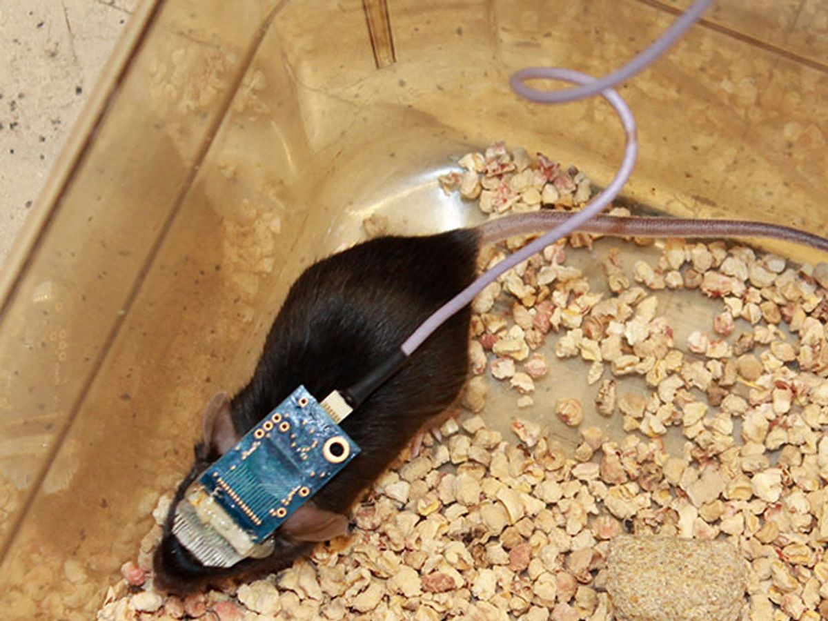
Now researchers at Harvard have shown that they can track brain activity, at the level of individual neurons, for months at a time, using a tiny electronic mesh that can be injected directly into the brain. A group led by Charles Lieber, a chemistry professor at Harvard, reports in this week’s Nature Methods that they were able to record the neural activity of mice over eight months, long enough to see how the animals’ brains changed as they entered the mouse version of middle age.
“This really brings us the ability to pick apart the circuits that are involved in fundamental information processing with the brain as they develop,” Lieber says.
The team builds the mesh out of very thin silicon wires coated in a polymer, with crossing lines made entirely of polymer; together they form simple field-effect transistors. The mesh naturally curls up when it’s put in a liquid, and can be drawn into a syringe and injected. Once in the brain, the mesh uncurls and sits on top of the neurons.
Implantable electrodes already exist, of course; doctors place them in the brains of some Parkinson’s patients to provide deep brain stimulation, which can help control tremors. But such devices are large and stiff and tend to irritate the brain tissue. The brain responds by engulfing them in a layer of cells, which insulates them and makes receiving or transmitting electrical signals more difficult.
By contrast, Lieber’s meshes are soft and flexible, and the transistors they form are smaller than the brain cells; they don’t provoke an immune response and they stay where they’re put. Where other implants lose their usefulness in days or weeks, the Harvard team’s meshes kept functioning for the entire length of the eight-month experiment.
The researchers also included some electrodes that could provide electrical stimulation to the brain. Lieber’s hope is that, if scientists can identify what goes wrong in the brain’s circuitry—leading to, say, Parkinson’s—at an early stage, they could use some sort of stimulation to alter or at least slow the process.
Statistical analysis of the signals they recorded showed that they were picking up activity from individual neurons, and that they could follow the same neurons over time. This ability could provide neurologists with a detailed map of what’s going on in, say, the visual cortex during learning, or let them watch the process by which memories are formed and how that process degrades with age.
Lieber would also like to use the devices in other parts of the nervous system. A mesh over the retina might yield some information about what’s happening in the eye and how that ties into what the neurons are doing. A set of electrodes in the spinal cord might provide new information, or even a new form of therapy, in cases of traumatic injury.
Neil Savage is a freelance science and technology writer based in Lowell, Mass., and a frequent contributor to IEEE Spectrum. His topics of interest include photonics, physics, computing, materials science, and semiconductors. His most recent article, “Tiny Satellites Could Distribute Quantum Keys,” describes an experiment in which cryptographic keys were distributed from satellites released from the International Space Station. He serves on the steering committee of New England Science Writers.


