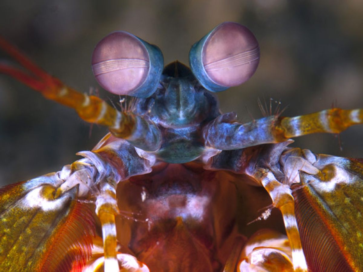
Millions of years of evolution have given the Mantis shrimp compound eyes to spot delicious meals that it can either spear or club to death in its underwater environment. More recently, the natural design of those eyes has inspired a new camera sensor that could spot cancer cells inside patients' bodies.
Engineers developed the sensor to mimic the Mantis shrimp's ability to filter polarized light—light waves vibrating along a single plane. Sensitivity to polarized light can help with cancer detection because cancerous lesions tend to reflect light in a way that causes more depolarization than does healthy tissue. Tests in mice have already shown how the new sensor can help detect flat, depressed, cancerous lesions that might otherwise be difficult to spot during a traditional endoscopy exam.
"Nature has [been] coming up with elegant and efficient design principles, so we are combining the mantis shrimp’s millions of years of evolution—nature’s engineering—with our relatively few years of work with the technology," said Justin Marshall, a neurobiologist at the Queensland Brain Institute of the University of Queensland, Australia, in a news release.
Mantis shrimp filter polarized light through their eye cells by using microvilli that resemble tiny hair-like objects. By comparison, the new sensor uses aluminum nanowires that together act as linear polarization filters that achieve a similar effect. Marshall and his international colleagues described the new sensor in a review article published in the journal Proceedings of the IEEE.
So far, it's the only polarization imaging sensor capable of fitting on the front top of the flexible endoscopes that physicians use to snake through patients' bodies to look inside. That could prove useful for helping to identify hard-to-see lesions that don't stand out from healthy tissue.
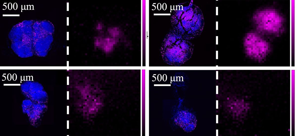
In the tests on mice, the sensor was used alongside a fluorescence-sensitive CCD camera that is designed to spot cancerous regions because a special fluorescent dye accumulates in abnormal tissue more than it does among groups of normal cells. Several members of the international research team, including engineers at Washington University in St. Louis, also developed high-tech goggles capable of helping surgeons detect cancer via such fluorescent markers.
The polarization sensor has other uses that go well beyond cancer screening. For instance, the Proceedings of the IEEE journal article laid out the vision for creating an implantable neural recording device that could help study neural activity in awake, freely-moving animals. It also pointed to the possibility of having real-time imaging that shows the stress and strain of soft biological tissue. Marine biologists at the University of Texas at Austin have even used the new sensor to study how female swordtail fish are attracted to ornamental patterns on the tails of male swordtail fish that are only visible in polarized light.
Jeremy Hsu has been working as a science and technology journalist in New York City since 2008. He has written on subjects as diverse as supercomputing and wearable electronics for IEEE Spectrum. When he’s not trying to wrap his head around the latest quantum computing news for Spectrum, he also contributes to a variety of publications such as Scientific American, Discover, Popular Science, and others. He is a graduate of New York University’s Science, Health & Environmental Reporting Program.


