It’s always a bit astonishing to discover that ancient technologies like the Lycurgus Cup achieved their seemingly miraculous effects because of nanoparticles. Strictly speaking, these examples are not “nanotechnology” because the engineers who developed them did not create them intentionally or even understand how they worked. Nevertheless, these examples underline how nanotechnologies can impact the products that are around us—in some cases for centuries.
The latest addition to this list of old technologies enabled by nanotechnology was the recent discovery that daguerreotypes are in fact made up of nanoparticles. The revelation occurred after curators at George Eastman House International Museum of Photography and Film in Rochester, NY, started to notice that several of the daguerreotypes in their museum were fading or had a white haze that obscured the images.
The preservation of these images is critical because daguerreotypes represent the first form of photography when Louis-Jacques-Mandé Daguerre invented it in 1839. In addition to their historical significance, the incredible resolution that can be achieved with these images could instruct future optical technologies.
When the conservators at the Eastman House were unable to explain why the degradation was happening, they called upon Nicholas Bigelow, a physicist at the University of Rochester to determine the cause. Bigelow confirmed earlier speculation that the damage to the images was being caused by fungi interacting with the surface of the daguerreotypes.
After discovering the cause, the Eastman House conservators started to store the images in argon gas to keep them in an a sort-of suspended animation to prevent the fungi from spreading until a more permanent solution can be developed. A video of this project can be seen below:
What may be even more significant about the work that Bigelow and his fellow researchers conducted is that it has shed further insight into the self-assembly of nanoparticles. By increasing control over the self-assembly of nanoparticles, researchers are developing better cancer treatments and a new generation of electronics.
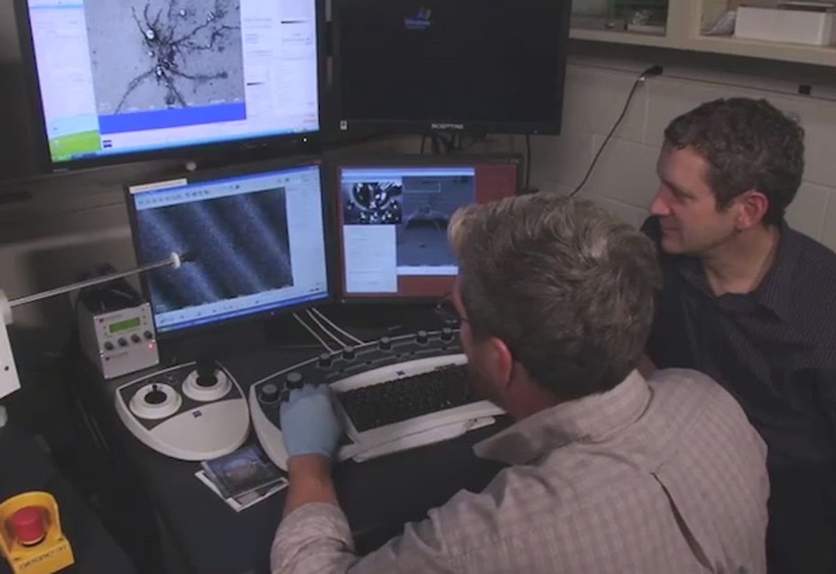
Bigelow and his research team employed an arsenal of microscopy tools, including a focused ion beam, a scanning electron microscope and a transmission electron microscope, to determine what lay beneath the nanoparticles. They discovered there were holes, pores, and cavities that were at least part of the problem with the degradation of the daguerreotypes. But what may be a bust for daguerreotypes could be a boon for other nanoparticle applications in areas such as medicine for medical capsules, according to Bigelow in the press release.
The truth is that despite the latest microscopy tools, the researchers at the University of Rochester still don’t fully understand the fundamental physics at work with the nanoparticles in the daguerreotypes. If they can solve that riddle, a more permanent solution to preserving the images and developing new applications for the nanoparticles could be reached.
“Nobody really robustly understands what’s happening, either to create the image or what’s happening as the image degrades,” says Brian McIntyre, a senior engineer on the project. “Understanding the fundamental chemistry and physics of the daguerreotype process is seminal to understanding how to preserve them.”
Dexter Johnson is a contributing editor at IEEE Spectrum, with a focus on nanotechnology.


