
A technology that better targets an X-ray imager’s field of view could allow various medical imaging technologies to be integrated into one. This could produce sharper, real-time pictures from inside the human body, says a researcher who hopes to one day build such a unified imager.
Ge Wang, the director of Rensselaer Polytechnic Institute’s Biomedical Imaging Cluster, in Troy, N.Y., calls his vision omni-tomography. Mixing and matching imaging techniques, such as computed tomography, magnetic resonance imaging, and single-photon emission computed tomography, could improve biomedical research and facilitate personalized medicine, says Wang, an IEEE Fellow.
To fit these imaging methods together, Wang and his collaborators have been developing a technology called interior tomography. In standard CT, X‑rays pass through two-dimensional slices of the body, and then a computer processes the data to build up a picture. If the scanner is trying to image the aorta, for instance, it will X-ray a whole section of the chest, including the points where the body ends and the open air begins. That boundary provides the image-building algorithm with defined edges and the background information it needs to operate. But interior tomography focuses only on structures inside the body, which reduces the patient’s radiation exposure. “If you’re only interested in the heart, why bother to cover your whole chest with X-rays?” says Wang.
Narrowing the view, however, eliminates the usual reference points needed to create an image conventionally. Interior tomography relies on a different set of hints. The new technique uses information about how substances within the body (such as blood) and air pockets alter X-rays to provide the algorithm with a base for reconstructing the image. It can even use old X-ray images of the same patient to help out.
Focusing on a specific region has advantages, particularly with patients too big for conventional scanners. “If an object is wider than the X-ray beam width, classic theory says you cannot do an accurate reconstruction,” says Wang. That’s not a concern with interior tomography, he says.
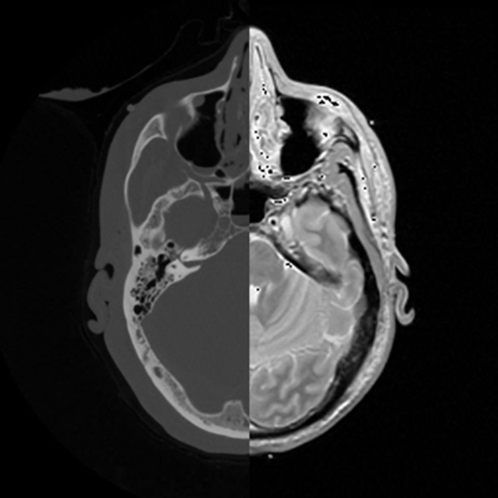 Seeing Both Sides: A CT scanner’s X-rays (left side) provide good structural detail; MRI images (right) are good for seeing functional flaws. Omni-tomography could combine the two technologies in the same machine.Images: Hao Gao & Ge Wang
What’s more, Wang’s team has shown that this concept can be generalized for use in imaging methods other than CT scanning, including MRI. And that could lead to a true fusion of major medical imaging techniques. In part that’s because the technique allows the use of smaller X-ray detectors, which in turn makes it possible to fit more scanners into the same machine.
There are already systems that combine two imaging methods—PET and CT or SPECT and CT, for instance. But those systems usually apply different methods in sequence rather than simultaneously, making it harder to see biological processes in action. The combination of CT and MRI has never been attempted before, but Wang says it’s possible now.
In fact, he and his collaborators in Australia, China, and the United States recently came up with a top-level engineering design for a CT-MRI scanner. They hope to present their design in June at the International Meeting on Fully Three-Dimensional Image Reconstruction in Radiology and Nuclear Medicine, in California. Applying interior tomography to MRI imaging allows the use of a weaker magnetic field, which is one way the design compensates for the incompatibility between powerful magnets in the MRI and rotating metal parts in the CT scanner.
Wang’s team does not yet have the funding to build a combination CT-MRI scanner, but putting the two technologies together could prove useful. MRI gives high contrast and allows doctors to measure functional and even molecular changes; CT provides greater structural detail. Together, they might allow doctors to get a superior picture of processes in action, such as changes during a heart attack, or serve as a guide to a surgical procedure. The technology would be ideal for imaging vulnerable plaques, suggests Michael Vannier, one of Wang’s collaborators and a radiology professor at the University of Chicago. Vulnerable plaques are buildups on artery walls that are particularly unstable and prone to causing heart attack or stroke. A combination of structural, functional, and molecular information is needed to tell just how dangerous the plaque may be. “In the long run, we think putting many imaging modes together will give you more information,” Wang says.
Interior tomography “is certainly an interesting concept that takes the interest in combining modalities to the ‘ultimate’ level of a single device,” says Simon Cherry, director of the Center for Molecular and Genomic Imaging at the University of California, Davis. While omni-tomography is technically feasible, Cherry wonders whether it will make sense from a clinical and economic perspective. “There are some that say too many of our health-care dollars are spent on imaging, especially in the pursuit of defensive medicine. This will be an expensive machine,” he says. “These are the issues that may well determine whether this approach is successful.”
About the Author
Neil Savage describes IEEE Fellow Ge Wang’s research on interior tomography, which he first came across while reporting on how medical device firms were trying to reduce the X-ray dosage produced by their imagers. Savage, a longtime Spectrum contributor, says, “With the growing popularity of scanning, finding ways to limit radiation exposure is important.”