
28 July 2010—The risk of getting cancer from CT scanners is still controversial in the medical community. But the equipment is being used more than ever: More than 70 million CT scans were performed in the United States in 2007. So scanner manufacturers and radiologists are devising new ways to reduce patients’ radiation exposure. Until now, the focus has been on software tricks that get cleaner images from smaller doses. But researchers at GE Global Research in Niskayuna, N.Y., and Stanford University have recently come up with a new scanner design that they say could reduce the needed radiation by up to 70 percent. Their design flips the conventional CT scanner design on its head.
Today’s machines contain one or two X-ray sources and a large arc-shaped detector opposite the sources. The sources and detectors revolve around the patient as a unit, taking multiple images that are put together to give a cross-section or 3-D picture of the body structure.
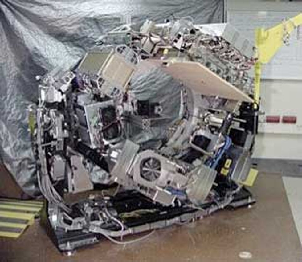
The new "inverse geometry" scanner will have tens of X-ray sources and a small detector with sensor elements that are just a fifth the size of today’s detectors. The sources, lined up in two rows, would rapidly and sequentially fire X-rays for just 10 microseconds each, covering a 16-centimeter span of the body four times larger than is possible for one conventional x-ray source. The researchers have tested a prototype that incorporates eight sources with standard GE scanner parts. They presented their results on 19 July at the American Association of Physicists in Medicine meeting and are now working on integrating all 32 sources.
In an X-ray source, a high voltage accelerates a focused electron beam across a glass tube. Electrons slam into a focused spot on the anode, and X-rays come out. Ideally, the rays pass through the patient in a straight line, hitting the detectors on the other side.
But the X-rays scatter. Conventional detectors have blades of tungsten or lead between adjacent detector cells to absorb and filter out the scattered photons. The detectors for the new distributed source do not need these antiscatter grids, allowing the researchers to squeeze in more sensor elements into a smaller area. "Every X-ray beam is very narrow, so we don’t have many scattered photons," says Bruno De Man, who is leading the work at GE. What's more, the intensity of each beam can be adjusted separately according to the patient’s anatomy, size, and weight, "so we can send X-rays only when and where we need them," he says. "We could avoid sensitive organs and really minimize [the] dose to the patient."
Generating multiple narrow X-ray beams was one of the challenges GE worked on. The researchers started with sources that had a so-called dispenser cathode known for generating an intense electron beam. But these are pretty inefficient. About 99 percent of the electrons hitting the anode generate heat instead of X-rays. The anode required cooling in the form of a substantial copper heat sink to reach 60 kilowatts (CT scans typically need 50 to 120 kW). By flowing oil through the anode for cooling, the team could increase the power to 100 kW.
Other challenges included keeping the electron beam focused to millimeter precision and designing the hardware to survive electrical discharges and ions created by the high voltage in the tube. The researchers also plan to halve the size of the detector and the spot that the beams focus on to get even sharper images, De Man says. He adds that the technology will not be mature enough for the market for at least another five years.
Manufacturers and researchers are already working to cut the radiation dose from existing scanners. Both GE and Siemens have incorporated a software technique known as iterative reconstruction, which reduces noise in images created by lower-intensity X-rays, effectively giving high-quality pictures with less radiation, says Cynthia McCollough, professor of radiological physics at the Mayo Clinic in Rochester, Minn. McCollough is working with a manufacturer she would not name on a way to automatically modulate the energy spectrum of the X-rays. Lower-energy X-rays are more sensitive to the density and composition of the material they are passing through and, for children and thinner adults, can give better image quality at 20 to 40 percent less radiation.
McCollough thinks the inverse geometry approach and other cutting-edge technologies such as photon-counting detectors are in their infancy but seem promising in the long run. The radiation dose of 1 to 15 millisieverts delivered by a CT scan is on a par with the average range of background radiation individuals receive in the United States every year—1 to 10 mSv—but there will always be room for technology improvements to reduce doses further, she says. "You always want to expose your body to the lowest dose that gets the job done."
About the Author
Prachi Patel is a contributing editor at IEEE Spectrum and a freelance journalist based in Pittsburgh, Penn. In the July 2010 issue she explained the computing behind the decoding of the Neanderthal genome.